Hi Everyone,
Great job this year on blogging our Honors Biology Class. I think that you all learned a lot this year and so did I. Have a wonderful summer and I'll see you in the fall.
Mrs. Andrews
Wednesday, June 8, 2011
Wednesday, June 1, 2011
June 1, 2011
Yo wat up.
we did a lot of notes today....NERVOUS SYSTEM!
Neuron- nerve cell specialized for carrying signals from one part of the body to another.
Nerve- the communication line made from bundles of neuron fibers wrapped in connective tissue.
The nervous system has two MAIN parts. The CNS (Central Nervous System) and the PNS (Peripheral Nervous System)
The functions of the N.S. are sensory output, Integration/Association interneurons, and Motor Output.
Motor Neuron
- has nucleus and other organelles
- Dendrites- branched, short, and recieve message from other cells
- Axon- long fiber that conducts signal toward another neuron or effector.

- Supporting cells- protect, insulate, reinforce
- Myelin sheath- chain of beadlike supporting cells
- Nodes of Ranview- spaces in myelin where impulse is transmitted
- synaptic knob- relays signals to other neurons
The signals move through the neuron in a weird way. In rest, the INSIDE IS NEGATIVE AND THE OUTSIDE IS POSITIVE. A stimulus is something that causes a nerve signal to start. Action potential is self-propagating change in voltage across plasma membrane.
How does the impulse move from neuron to cell?
- Electrical Synapse
 action potential jumps from cell to cell
- heart and digestive track
- Chemical synapse
- synaptic cleft- narrow gap seperating 2 cells.
- most other organs, skeletal muscle and CNS
Neurotransmitter- chemical carrying info from neuron to another type of cell that will react
- Action Potential arrives at the synaptic knob
- Neurotransmitter vessicals fuse with membranes
- neurotransmitters are released into cleft
- neurotransmitter diffuse across gap and binds to receptor proteins on receiving neurons
- Ion channels open and ions trigger action potential.
- Neurotransmitter are broken down and returned to sending neoron for recycling
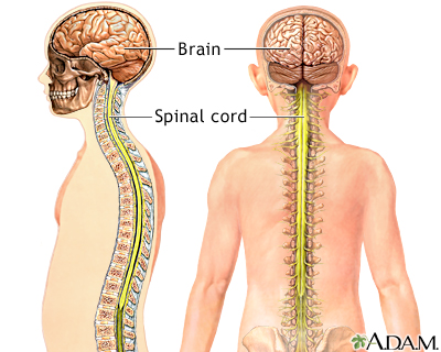
CNS
- brain and spinal cord
- Cephalization- concentration of nervous system at the head end.
- Centralization- presence of CNS saparate from PNS
- spinal cord- bundle of nerve fibers in spinal column (Central communication between brain and body)
- Brain- Master control center (Homeostasis center, sensory center, emotion, intellect
- Brain as cerebrospinal fluid - liquid that cushions the CNS and helps supply it with nutrients, WBC, and hormones.
- Has Meninges- layers of connective tissue for protection
- white matter- mainly axons (with myelin sheath)
- gray matter- mainly nerve cell bodies and dendrites (Cerebral Cortex)
PNS
- Has a couple of divisions.
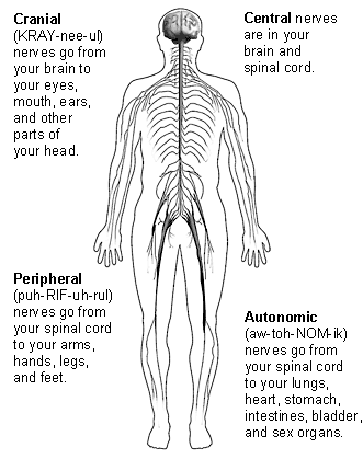
- Sensory Division
- brings info to CNS from outside environment
- Info into CNS from body itself (internal environment)
- Motor Division
- Somatic Nervous System- carrys signals to Skeletal muscles (voluntary, concious control)
- Autonomic Nervous System- controls smooth and cardiac muscle, organs, glands, digestive, circulatory, excretory, and endocrine (2 branches and involuntary)
Yep! Thats all folks!
HW:
- Study for finals
- UP pg. 81-84
- study for test Mon.
- Crossword puzzle
Tuesday, May 31, 2011
5/31/11
What we did today:
- Notes
- Balloon lab and Lab #65: UP 57-61
Smoking
- Air pollutants: CO, SO2, O3
- It contains more than 4000 chemicals
- The chemicals damage the mucus and cilia making it difficult to remove foreign particles
- It kills over 430,000 Americans per year
- Emphysema: disease that causes alveoli to disintegrate causing breathlessness and fatigue

Thursday, May 26, 2011
Respiration
5/26/11
Yunsu Y.
Respiration notes:
** The respiration that we are talking about is not the same as cellular respiration!
1. Respiratory Surfaces:
** not all organisms have respiratory systems: bacteria don't have because their surface area to volume ration is large so diffusion can occur, but for humans our surface area is too small so we need respiratory systems.
 Aveoli close up
HW: Study for Fetal Pig Dissection Test TOM!!
Lab 54&55 due tom with cover page and color code the pictures
U.P 47-51 and 55-56 due TUESDAY
Next Scribe CJ P.
Yunsu Y.
Respiration notes:
** The respiration that we are talking about is not the same as cellular respiration!
1. Respiratory Surfaces:
** not all organisms have respiratory systems: bacteria don't have because their surface area to volume ration is large so diffusion can occur, but for humans our surface area is too small so we need respiratory systems.
- Earthworm: has moist skin=making diffusion across body surface easier.
- Aquatic Organisms: gills extend from the body which increases surface area and they are surrounded by water so that diffusion can occur.
- Terrestrial Organisms: respiratory surfaces fold into the body. The system is inside lined with moisture (mucus), needed for diffusion.
- Insects: tracheae and no circulatory system to transport O2.
2. Human Respiratory System:
- Breathing: the moist insides are exposed to air, then O2 dffuese into blood vessels and CO2 to the lungs.
- Circulatory System: trasnport O2 to all body cells and O2 back to lungs.
- Diaphragm: Sheet of muscle-important for breathing.
- Pharynx: where the digestive and respiratory systems meet.
- Larynx: voice box, we produce sounds by breathing out and the air moves the vocal cords making them vibrate.
- Trachea: windpipe, somtimes food gets caught here causing us to cough or choke.
- Bronchi: Lead to each lung
- Bronchioles: smaller branches
- Alveoli: cluster of air sacs. These are like the villi and microvilli found in the small intestine. This is where gas exchange occurs with blood vessels. These increase the surface area=more gas exchange.

Aveoli close up3. Taking a Breath
- When the diaphragm contracts air is "PULLED" into the lungs. This is increasing the volume and decreasing the pressure. High pressure outside wants to get inside to the low pressure. (high to low!)--------- Negative Pressure breathing.
- When diaphragm relaxes when the air is "PUSHED" out of the lungs. There is a decrease in volume and an increase in pressure.
- Nerves in the brain regulate our breathing. Nerves tell the diaphragm when to contract and relax.
- 10-14 inhalations per minute. (average)
- This average will change depending on CO2 levels. The more CO2 in the blood, the faster the respiration rate.
- Hyperventilation: Purges blood of CO2 that brain stops sending messages to the diaphragm. This causes people to pass out but breathing into a paper bag helps by increasing the amount of CO2 taken in.
- Consists of 4 polypeptide chains, heme (chemical group) and iron- gives blood the red color.
- Each iron atom can bind to 1 O2 molecule=each hemoglobin can carry up to 4 O2 molecules.
- O2 rich blood is bright red and O2 poor blood is dark red or blue.
- Iron deficiency cause anemia
- When hemoglobin binds to CO (carbon monoxide)=Bad! Now the hemoglobin cannot bind with oxygen because the bond is too strong on the CO. This interferes with the delivery of O2 to the body cells and cellular respiration=death. Also found in cigarette smoke. \
- Pollutes air
- Contains many harmful chemicals damaging mucus and cilia making it hard to remove foreign particles. = smokers cough.
- Smoking kills ~430,000 Americans per year.
- Emphysema: disease that causes alveoli to disintegrate. = reduces lungs' ability to exchange gas and causes breathlessness and fatigue.
Lab 54&55 due tom with cover page and color code the pictures
U.P 47-51 and 55-56 due TUESDAY
Next Scribe CJ P.
Wednesday, May 25, 2011
Pig Dissection Day 3
Heart and Lungs Continued:
We used scissors to cut the blood vessels around the heart. After examining the heart and the vessels, we examined the back, or dorsal, side of the heart. Afterwards, the heart was cut lengthwise (the cut was parallel to the front and back of the heart). Both ventricles and the left atrium were visible inside of the heart. With the heart removed, we identified the trachea and esophagus. The trachea is the windpipe that carries air. The esophagus is the tube that carries food, liquids, and saliva from your mouth to stomach. The carotid arteris and jugular veins, which carry blood to and from the head, were found on either side of the trachea. Beneath them was the vagus nerve. The thyroid gland was placed ventral to the trachea. It was reddish-brown and had two lobes. The larynx was located at the top of the trachea.
The Head:
We opened the mouth of the pig and examined the tongue, any teeth visible, and the back of the throat. We had to use our scalpel to slit the corners of the mouth on both sides in order to view the epiglottis, glottis, and opening to the esophagus. The pig has four pairs of salivary glands; the largest of these is the parotid gland, which extends from the base of the ear to the shoulder and the jaw. Using our scalpel, we made an incision through the skin and facial muscles at the base of the ear. The skin and muscle layer were removed, and we examined the parotid gland. Beneath the parotid gland was the mandibular gland.
The Nervous System:
The pig has a very similar nervous system to that of humans. There is the central nervous system consisting of the brain and spinal cord, and the peripheral nervous system consisting of cranial and spinal nerves and their branches. Using our scalpel, we made an incision through the skin of the head. The skin was then peeled off. Afterwards, we inserted the pointed end of our scissors between the area where the bones of the skull met. Then we broke off pieces of the skull until most of the skinned area was open. The outermost membrane of the pig is called the dura mater. It is the thickest and toughest of the membranes. The surface of the brain is covered by a thin membrane called the pia mater. The third membrane is called the arachnoid membrane and is found between the dura mater and pia mater. In living animals, cerebrospinal fluid fills the space between the two inner membranes. Our group frist cut through the dura mater, exposing the brain. We attempted to identify the right and left cerebral hemispheres, cerebellum, cerebrum, olfactory lobes, and the medulla. We also identified cranial nerves. The spinal cord was surrounded and protected by the vertebrae of the spinal column. We removed the skin from an area of the back so around 8~9 cm of the spinal column were exposed. The remaining tissues were removed so the spiny extensions of the vertebrae were not completly exposed. We cut off the tops of the spiny extensions of the vertebrae with our scissors and saw the spinal cord and spinal nerves.
HW: Lab with cover sheet due Friday! (Don't forget to color code diagrams in lab).
Next Scribe: YUNSU Y. (:
Monday, May 23, 2011
Pig Dissection Day 1
FETAL PIG DISSECTION: Day 1
We started on our first incisions today after we tied down our pigs. The tying helped make clean cut incisions. These incisions led to seeing the abdominal cavity of the fetal pig. The liver takes up most of the upper part with the lungs and the intestines taking up most of the bottom. The gallbladder is hidden behind the lungs and the stomach is the balloon like structure kind of under the liver.
The sex of the pig can be determined by looking underneath the anus. Both males and females have nipples on the ventral surface. The females have a spike-like genital papilla. Males have a scrotal sac containing the testes under their anus.
Dissection to be continued in the next few posts.
Next scribe: Sally Y. :]
Sunday, May 22, 2011
May 20th 2011

CJ P
White blood cells v. Platelets
WHITE BLOOD CELLS:
aka: leukocytes
WBCs fight infection
Lack hemoglobin (which is found in RBCs)
Have nucleus (not found in RBCs)
Found OUTSIDE of circulatory system
PLATELETS:
aka: thrombocytes (bits of cytoplasm broken off from bone marrow)
Fibrinogen: protein found in plasma
Fibrin: Fibrinogen converted by clotting
CLOTS BLOOD (scabs)
HEMOPHILIA: excessive, fatal, bleeding occurs from minor cuts
THROMBUS: blood clot that forms in absence of injury
EMBOLUS: thrombus that dislodges and travels
Cardiovascular disease: set of diseases that affect the heart.
The heart requires oxygen rich blood to survive because it's made of TISSUE.
CORONARY ARTERIES: supply heart muscle with oxygen
HEART ATTACK: failure of heart to function properly
ATHEROSCLEROSIS: chronic cardiovascular disease
Cholesterol build up in arteries causing narrow passages--->decreased blood flow----> increased blood pressure= BAD.
TREATMENTS:
ANGIOPLASTY: baloon catheter to compress plaque=opens clogged arteries
STENT: wire mesh tubes prop open arteries
BYPASS SURGERY: vessel sewn onto heart shunt blood around a blocked artery
ARTIFICIAL HEART: an artificial heart.
lots of homework:
Pgs 7-18
Lab 15
Read pig lab -closed toed shoes and hair ties required in case fetal pigs turn into zombies, you will be prepared to run.
43-46
Next scribe: Smally Patty (Sonali)
Subscribe to:
Posts (Atom)